The central concept: Fractures fail to unite when their mechanical environment, local biology, infection status or patient factors are unfavorable. Correcting only one element may not be enough.
1. Delayed union, nonunion and malunion: what is the difference?
Most fractures progressively develop stable bony union. When healing is slower than expected, the clinician must examine serial images alongside pain, mechanical stability and the patient's biological health.
| Term | Practical meaning | Key distinction |
|---|---|---|
| Delayed union | Healing is taking longer than expected for the specific bone, injury and fixation method. | Progress may still occur; review serial films and causes. |
| Nonunion | Fracture repair has arrested or is highly unlikely to proceed without a change in treatment. | Persistent pain/instability and failure of radiographic progression. |
| Malunion | Fracture has healed but in an unacceptable position. | Union may be solid despite deformity. |
2. Types of nonunion — the X-ray tells a mechanical story
Two useful classifications overlap: biological/radiographic morphology (hypertrophic, oligotrophic, atrophic) and infection status (aseptic or infected). Never assume morphology alone excludes infection.
 ↗ Click to enlarge
Hypertrophic nonunion — plenty of callus, no bridge
Radiographs show abundant callus around an ununited site (“elephant-foot” appearance). Healing biology is often active, but excessive motion or instability prevents successful bridging.
Usual treatment principle: improve stability and correct alignment; grafting is not automatically required.
 ↗ Click to enlarge
Atrophic nonunion — sparse callus
Little visible callus, persistent gap and sometimes sclerotic ends suggest impaired healing biology and/or unfavorable mechanical conditions. Avascularity must not be diagnosed from an X-ray alone.
Usual treatment principle: restore stability, correct alignment, remove nonviable tissue where appropriate and optimize biological support.
| Pattern | Typical appearance | Management emphasis |
|---|---|---|
| Hypertrophic | Abundant periosteal callus without continuity | Often primarily mechanical correction |
| Oligotrophic | Little callus, often poor fragment contact or displacement | Improve alignment/contact and stability; assess biology |
| Atrophic | Minimal callus, narrow/rounded or sclerotic fracture ends | Mechanical correction plus biological optimization as needed |
| Infected | Variable morphology; may show lucency, sequestration or loose implants | Source control + stability + tailored antimicrobial treatment |
3. Why fractures do not heal
Most nonunions have more than one contributing factor. Organize the causes in four groups; this makes clinical evaluation and treatment far easier.
| Mechanical / injury | Biological / patient |
|---|---|
| Instability, gap, distraction, malalignment, poor construct, major comminution, segmental loss | Severe soft-tissue damage, disrupted vascularity, smoking/nicotine, diabetes, malnutrition, vitamin D deficiency where relevant |
| Open or high-energy injury, repeated surgery, loss of cortical contact | Infection, immunosuppression, metabolic/endocrine abnormalities, some medication exposures |
Fixation construct problems
Plate breakage or loosening may indicate continuing cyclic load across an unhealed fracture. The reason for construct failure matters: persistent fracture gap, varus deformity, overly concentrated stress or infection may all contribute. Do not simply replace a broken plate with the same construct without addressing the underlying cause.
4. Clinical presentation and diagnostic workup
 ↗ Click to enlarge
Why serial imaging matters
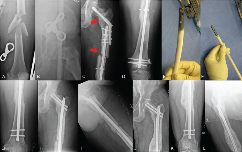
This published case illustrates a femoral neck–shaft nonunion and varus malalignment followed by revision management. Compare fracture contact, neck-shaft alignment, hardware integrity and interval progression rather than judging one isolated film.
CT supplements radiographs; it does not replace clinical assessment or microbiological investigation.
5. Always consider fracture-related infection (FRI)
An apparently “aseptic” nonunion may conceal infection. Use the international FRI consensus distinction between confirmatory and suggestive features.
| Confirmatory findings | Suggestive only |
|---|---|
| Sinus/wound breakdown communicating with bone or implant; pus; matching pathogen in ≥2 separate deep specimens; specified histopathological findings | Redness, warmth, swelling, pain, persistent drainage, loose implants/osteolysis, elevated CRP or ESR, a single positive deep culture |
 ↗ Click to enlarge
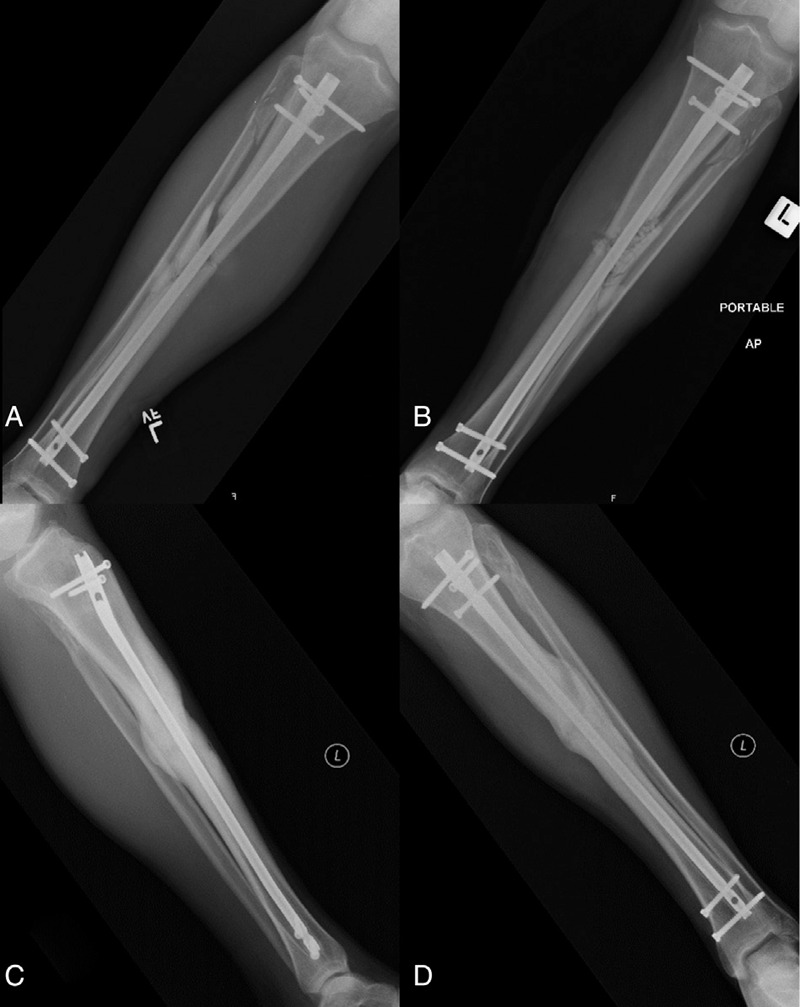
Real tibial infection-to-union sequence
Four radiographic panels from a published case show acute infection after tibial nailing, antibiotic treatment while stable implants were retained, and later union. Implant retention is selective—dependent on stability, soft tissues, infection timing and feasibility of adequate debridement.
In chronic established infected nonunion, revision strategies frequently require implant exchange or removal, debridement and restabilization.
6. Treatment — correct the cause, not just the X-ray
Choose treatment after identifying whether the site is stable, biologically viable, aligned, infected and functionally problematic. A selected progressing delayed union may justify observation and risk-factor correction; established symptomatic nonunion often requires intervention.
Deep sampling, debridement, source control, stability, pathogen-directed therapy
Correct instability/deformity/gap; improve biology as indicated
Nonoperative options — only in selected patients
Optimize smoking cessation, nutrition and diabetes; correct important deficiencies and review the fixation/loading plan. Protected weight-bearing, functional bracing and selected stimulation modalities may be considered where appropriate. Bone stimulators cannot compensate for gross instability, major distraction or untreated infection.
Surgical strategy depends on the failure mechanism
| Problem | Typical corrective strategy |
|---|---|
| Hypertrophic shaft nonunion with unstable fixation | Improve stability/compression and correct alignment, with grafting selectively |
| Atrophic/oligotrophic nonunion with defect or poor contact | Restore alignment/stability; consider autograft or biologic augmentation, address dead bone |
| Suitable aseptic femoral/tibial shaft nonunion after nail | Selected reamed exchange nailing, augmentation plating or revision construct depending on location and deformity |
| Infected nonunion | Culture-based debridement strategy, stable fixation, soft-tissue coverage and organism-directed antibiotics |
| Critical segmental bone defect | Masquelet induced membrane, bone transport or other specialist reconstruction |
 ↗ Click to enlarge
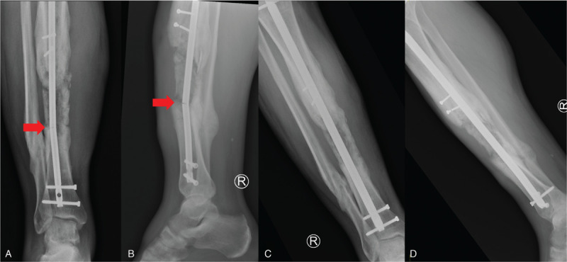
Exchange nailing and stability
This published tibial case shows failure of an intramedullary nail before the defect consolidated and subsequent exchange to a larger nail.
Exchange nailing can improve alignment/contact and stability in appropriately selected diaphyseal nonunions, but deformity, infection, large defects or metaphyseal geometry may favor another construct.
 ↗ Click to enlarge
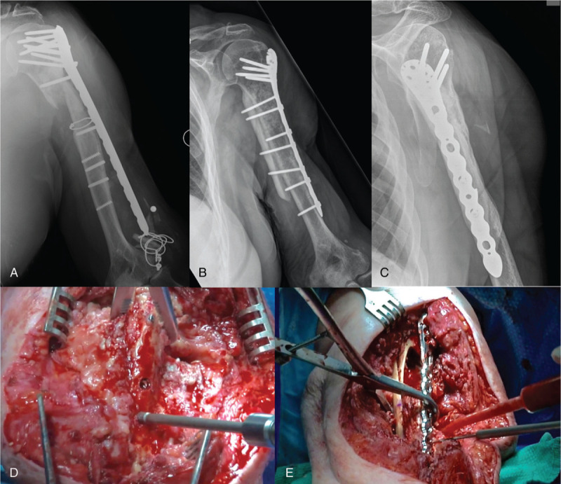
Revision plating and biological reconstruction
A proximal humeral nonunion with failed implants illustrates why revision requires planning for hardware removal, alignment, local biology and secure fixation.
Autologous cancellous bone graft offers osteogenic, osteoinductive and osteoconductive properties, but cannot replace adequate mechanical stability.
7. Advanced reconstructive options
Large defects, repeated treatment failure and infection require subspecialist planning. Advanced techniques are best understood as solutions to a specific problem—not interchangeable operations.
 ↗ Click to enlarge
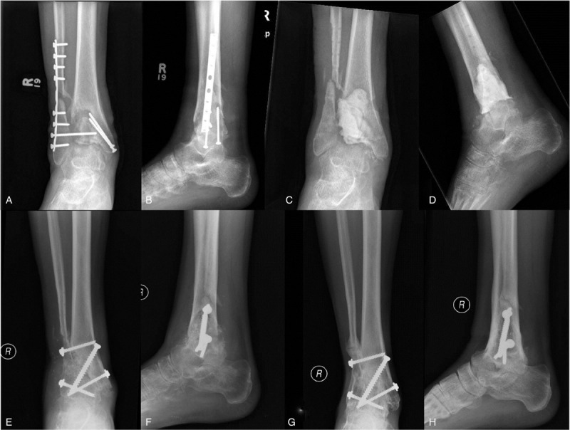
Masquelet induced-membrane technique
Stage 1: radical indicated debridement, stability, management of infection/soft tissues and insertion of a PMMA spacer into a segmental defect.
Stage 2: once conditions are suitable, open the induced membrane, remove the spacer, fill with bone graft and maintain stable fixation. Timing is individualized; “6–8 weeks” is often taught but is not mandatory in every patient.
 ↗ Click to enlarge
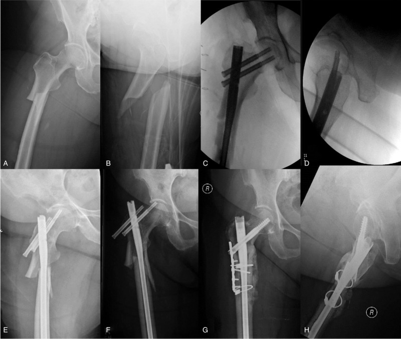
What if deformity remains?
A complex atypical femoral fracture series illustrates progressive varus collapse, failure of fixation and later revision. Do not graft an unstable varus construct without correcting its mechanical problem.
Revision may require correction of alignment, improved nail entry/trajectory, supplementary plating or other tailored fixation.
Optional advanced learning: bone transport, vascularized grafts and the diamond concept
Bone transport with circular or monolateral external fixation can reconstruct long defects through distraction osteogenesis; anticipate pin-site care, docking-site issues, joint contracture and prolonged treatment. Vascularized fibular grafting is a specialized option for selected large defects. The “diamond concept” integrates osteogenic cells, osteoconductive scaffold, growth mediators, vascularity and mechanical stability, within an appropriate host environment.
8. Special situations and common complications
 ↗ Click to enlarge
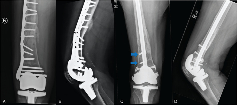
Periprosthetic distal femoral nonunion
This series demonstrates an unreconstructed/nonunited distal femur with plate failure and subsequent revision. Bone stock, prosthesis stability, alignment and fixation options dictate treatment; revision arthroplasty or distal femoral replacement may be considered in selected patients.
| Special location | Additional consideration |
|---|---|
| Femoral neck | Assess femoral head viability, varus, vertical shear and arthroplasty versus joint-preserving reconstruction. |
| Scaphoid | Review proximal pole vascularity, humpback deformity and possible need for graft/fixation. |
| Tibia after open fracture | Soft-tissue coverage, infection and segmental defect often dominate treatment. |
| Periprosthetic fractures | Check implant stability and available bone stock before choosing fixation versus revision. |
 ↗ Click to enlarge
Nonunion can occur at the wrist too
These real radiographs show distal radius/ulna pseudarthrosis after a wrist fracture. This is a site-specific example rather than a typical shaft nonunion: distal fragment size, joint alignment, bone stock and adjacent soft tissues change the fixation strategy.
Always interpret previous injury/fixation history before planning reconstruction.
9. Key concepts — 60-second recap
10. Interactive clinical viva — tap to reveal
Answers are hidden by default. Each item opens independently.
1. What distinguishes delayed union from established nonunion?
2. Does a hypertrophic nonunion need bone graft in every case?
3. Can normal CRP exclude infected nonunion?
4. How many cultures should be obtained during revision for suspected FRI?
5. What if an IM nail has failed in a tibial shaft nonunion?
6. What are the two stages of Masquelet?
7. Why do we get a CT scan?
8. Why does an atrophic nonunion need more than graft alone?
9. What defines FRI at the bedside?
10. How should periprosthetic nonunion be approached?
11. Interactive 20-question knowledge test
One question at a time. Select A–D for instant marking and option-by-option teaching feedback, then press Next Question. Saved locally in this browser; exam scores are not transmitted to a server.
Select an option to see the answer and unlock Next Question.
12. References & image credits
Show references and image credits (+)
Clinical reference sources
- Orthobullets — Nonunion and Bone Defects
- AO Foundation — Aseptic nonunion reference collection
- AO Surgery Reference — Nonunion management around knee implants
- Hoit, Bonyun, Nauth (2020). Hardware considerations in infection and nonunion management. OTA Int. CC BY 4.0
- Hak (2020). The infected fracture: can we agree on standard definitions? CC BY 4.0
- McNally et al. (2020). Definition and diagnosis of fracture-related infection.
- Metsemakers et al. General treatment principles for fracture-related infection.
- AAOS OrthoInfo — Nonunions
- US FDA safety/effectiveness document — nonunion definition
- AO — Core principles of fracture management
Images — direct original URLs and usage licences
Each figure links to its publication or Commons licence page. Images are displayed without cropping or editorial annotations; resized by CSS only. Some sources may restrict third-party hotlinking, so preview images on the published page.
- Atrophic distal humeral nonunion — AP and lateral radiographs
Attribution and licence: Hellerhoff / Wikimedia Commons (CC BY-SA 4.0)
Direct image URL - Tibial and fibular pseudarthrosis with abundant bony reaction
Attribution and licence: Jmarchn / Wikimedia Commons (CC BY-SA 3.0)
Direct image URL - Distal radius and ulna pseudarthrosis following a wrist fracture — two clinical radiographs
Attribution and licence: Jmarchn / Wikimedia Commons (CC BY-SA 3.0)
Direct image URL - Acute fracture-related infection and subsequent tibial union (four X-ray panels)
Attribution and licence: Hoit, Bonyun & Nauth / OTA International (2020), Figure 1 (CC BY 4.0)
Direct image URL - Femoral neck–shaft nonunion, varus malalignment and revision (multi-panel clinical series)
Attribution and licence: Hoit, Bonyun & Nauth / OTA International (2020), Figure 2 (CC BY 4.0)
Direct image URL - Atypical femoral fracture and revision for failed healing and alignment
Attribution and licence: Hoit, Bonyun & Nauth / OTA International (2020), Figure 3 (CC BY 4.0)
Direct image URL - Infected ankle nonunion treated with an induced-membrane (Masquelet) strategy
Attribution and licence: Hoit, Bonyun & Nauth / OTA International (2020), Figure 4 (CC BY 4.0)
Direct image URL - Proximal humeral nonunion with failed implants and revision fixation
Attribution and licence: Hoit, Bonyun & Nauth / OTA International (2020), Figure 5 (CC BY 4.0)
Direct image URL - Tibial nonunion with failed intramedullary nail and exchange fixation
Attribution and licence: Hoit, Bonyun & Nauth / OTA International (2020), Figure 6 (CC BY 4.0)
Direct image URL - Periprosthetic distal femoral nonunion, plate failure and revision fixation
Attribution and licence: Hoit, Bonyun & Nauth / OTA International (2020), Figure 7 (CC BY 4.0)
Direct image URL
Original educational synthesis written for FreeMedSite. This guide does not replace individualized orthopaedic assessment. Last content preparation: October 2026.


0 Comments